Precision oncology isn’t about better answers. It’s about asking better questions.

Progress rarely replaces previous knowledge. It builds on it.

Over the past two decades, our understanding of breast cancer has changed dramatically. What was once considered a single disease is now recognised as a collection of biologically distinct conditions, each with its own characteristics, behaviour and response to treatment. Every advance has revealed greater complexity, but that complexity has also created new opportunities for more personalised care.

As our understanding has evolved, so too have the questions researchers and clinicians ask.

Twenty years ago, the question was often, What type of breast cancer is this? Today, it is equally important to ask, What is this tumour doing now? How is it changing? Why does one patient respond differently from another? These questions reflect a shift from describing cancer to understanding its biology in real time.

This evolution is central to precision oncology.

Precision oncology is often associated with targeted therapies or genomic testing, but its broader goal is much simpler: to provide the right treatment for the right patient at the right time. Achieving that goal depends on understanding the biological signals that matter most at each stage of the cancer journey.

No single test can answer every question. Histopathology transformed cancer diagnosis by allowing tumours to be examined at the cellular level. Immunohistochemistry enabled more accurate tumour classification through hormone receptor and HER2 testing. Genomic profiling added another dimension by identifying molecular level alterations that influence prognosis and treatment selection. Together, these advances have become central to modern breast cancer management and are reflected in current international clinical guidelines.[1,2]

More recently, liquid biopsy has introduced the possibility of assessing tumour biology through a blood sample. Rather than replacing tissue biopsy, liquid biopsy offers an opportunity to complement established diagnostic approaches while enabling biological information to be collected over time.
Importantly, liquid biopsy is not a single technology.

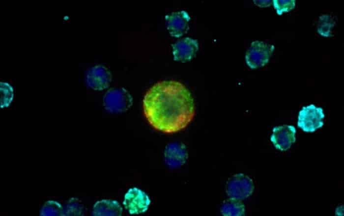
Circulating tumour DNA (ctDNA) consists of fragments of tumour-derived DNA released into the bloodstream and can provide information about genetic alterations. Circulating tumour cells (CTCs), by contrast, are intact cancer cells that have entered the circulation from a primary or metastatic tumour. Because they remain whole cells, CTCs may offer opportunities to investigate cellular characteristics, protein expression and tumour biology which complement ctDNA analysis.

This distinction highlights an important principle in precision oncology. Different technologies answer different biological questions.
Current evidence suggests that no individual biomarker can provide a complete picture of a patient’s disease. Instead, imaging, tissue pathology, genomic analysis, ctDNA and CTCs each contribute complementary information. Choosing the most appropriate mix of tools depends on the biological questions being asked.

This increasingly nuanced view is reflected in recent clinical guidance. The American Society of Clinical Oncology (ASCO) recognises the potential of circulating tumour DNA while emphasising that evidence supporting routine clinical implementation continues to evolve. Similarly, discussions at the 2026 ESMO Breast Cancer Congress highlighted the growing role of liquid biopsy across the breast cancer pathway, alongside the importance of rigorous validation for emerging biomarkers. [3,4]

As breast cancer treatment becomes more personalised, understanding tumour biology throughout the disease journey is becoming increasingly important. Tumours evolve. Treatments exert selective pressures. Metastases may develop distinct biological characteristics. These changes mean that the information needed at diagnosis may not always be the information needed months or years later.

This is where newer technologies are beginning to ask different questions.

Research into circulating tumour cells, for example, is exploring whether analysing intact cells may provide insights into tumour heterogeneity, treatment response and disease progression beyond genomic information alone. Although this work remains an active area of investigation, it reflects a broader trend across cancer research: recognising that understanding biology requires multiple complementary sources of evidence rather than a single definitive test.[5]
At Frontier, this principle underpins our developing work; to investigate circulating tumour cell technology as a means of supporting future research into tumour biology and disease monitoring. Our work is not CE-IVDR marked, UKCA approved, or FDA cleared and not available for clinical or commercial use. Like all emerging biomarker technologies, its potential must be established through robust scientific evidence before any future clinical application may be considered.

The history of breast cancer research shows that progress has rarely come from finding one perfect answer. Instead, each scientific advance has enabled researchers to ask better questions, revealing new layers of biological complexity and creating opportunities for more informed clinical decision making.
Precision oncology is not simply about developing new technologies. It is about understanding which questions those technologies are best placed to answer. As research continues, the greatest advances are likely to come not from relying on a single biomarker, but from combining multiple sources of biological information to build a more complete understanding of each patient’s disease.

Website disclaimer
Frontier Diagnostics’ technology platform currently in development. It is not CE-IVDR marked, UKCA approved, or FDA cleared. It is not available for clinical or commercial use. For research collaboration enquiries only.

Sources

1. Loibl S, Andre F, Bachelot T, Barrios C H et al. Early breast cancer: ESMO Clinical Practice Guideline for diagnosis, treatment and follow-up. Annals of Oncology.
2024
2. National Institute for Health and Care Excellence (NICE). Early and locally advanced breast cancer: diagnosis and management (NG101).
3. American Society of Clinical Oncology (ASCO). Circulating Tumour DNA Testing in Solid Tumours and Lymphoma: ASCO Guideline. 2026.
4. ESMO Breast Cancer Congress 2026. Liquid biopsy across the breast cancer spectrum.
5. Thomas-Bonafos T, et al. Emerging roles of circulating tumour cells in breast cancer research. npj Breast Cancer. 2024.

De-escalation – it sounds scary but……..

When less treatment may be the right treatment

De-escalation sounds scary to anyone who’s going through or has been through breast cancer. So why is the oncological world using the term? It sounds like care is being reduced – full-stop. The reality, however, is just the opposite – but also increasingly complex.

In breast cancer, the aim is to look at two types of information;

– the evidence building up from recent clinical trials as to when a treatment might not be effective for some people;
– better information becoming available on the individual patient’s needs.

De-escalation asks whether all this information can help clinicians identify when a less intensive treatment pathway is appropriate, without losing the cancer control that matters to patients.

Why the word can feel alarming

For many people, a breast cancer diagnosis creates an understandable instinct to do everything possible. More treatment for most patients feels safer – as if the more treatment we throw at it, the greater the chance of getting through it. The word de-escalation sounds as if care is being withdrawn, or that someone is being offered less because of pressure on services.

That is not the purpose of planned treatment de-escalation. It is an evidence-led approach that asks whether some people, can safely avoid treatment that is unlikely to add meaningful benefit. What de-escalation is about is using all the evidence from clinical research and the patient themselves to identify the optimum treatment regime and to give the right amount of treatment – no more, no less – to achieve the best results.

This is different from stopping, pausing or changing treatment because the side effects are too difficult to manage. A change made because someone is not tolerating treatment is an individual clinical decision.

Planned de-escalation is based on research evidence, better access to detailed information on tumour stage and size, tumour biology and biomarkers, response to neoadjuvant therapy, lymph node status as well as the patient’s age, general health and preferences.

Let’s look at a couple of examples of recent clinical research;

OPTIMA: what the first results showed

The first results from the phase III OPTIMA trial have brought de-escalation into public discussion. OPTIMA was designed to test whether a tumour gene expression test could guide chemotherapy decisions for people with early breast cancer who would traditionally be considered at higher clinical risk.[1,2]

The trial enrolled 4,429 women and men aged 40 or older with hormone-sensitive, HER2-negative early breast cancer. Most participants had cancer spread to nearby lymph nodes, which would usually make chemotherapy part of the standard discussion after surgery.[1]

Participants were randomly assigned to standard treatment, with chemotherapy followed by hormone therapy, or to a test-directed pathway using the Prosigna tumour profiling test. In the test-directed group, those with a low risk of recurrence score received hormone therapy without chemotherapy. Radiotherapy and other treatment were given as usual in both groups.[1]

More than two thirds of participants, 68%, had a low Prosigna score.[1,3] Among this low score group, five years after treatment, 95% of those who received chemotherapy plus hormone therapy were alive and free from breast cancer recurrence. The corresponding figure for those treated with hormone therapy alone was 94% in the UCL reporting of the trial.[1]

These results do not mean chemotherapy is unnecessary for everyone with hormone-sensitive breast cancer. They show that, in the population studied, additional tumour biology information helped identify many people for whom chemotherapy appeared to add little or no extra benefit.
They also show why de-escalation should never be framed as simply doing less. In OPTIMA, clinicians were not relying only on traditional features such as tumour size and lymph node involvement. They were adding biological information from the tumour itself to refine the decision.

More information is changing the question

Historically, treatment decisions have drawn on information such as tumour size, grade, lymph node involvement, hormone receptor status, HER2 status, menopausal status, age, other health conditions and patient preference. These remain essential.

What is changing is the amount and type of information that can now be considered. Tumour profiling can help estimate recurrence risk and likely treatment benefit in specific settings. Pathology continues to become more detailed. Imaging can help define disease extent and response. Blood-based approaches are being studied for signals of molecular change before those changes are visible on scans or symptoms are present.

This does not make treatment decisions automatic. It makes them more evidence-dependent. The more information clinicians use, the more important it becomes to know exactly what has been validated, in which patient group and for which decision.

Emerging biomarker and monitoring approaches

Biomarkers are often discussed as though they are a single category. In practice, different biomarkers answer different questions.
A tumour gene expression test, such as the test used in OPTIMA, is performed on tumour tissue and can help estimate risk or likely benefit from chemotherapy in a defined early breast cancer setting.[1,2]

Circulating tumour DNA, or ctDNA, is a blood-based signal made up of fragments of tumour-derived DNA. In breast cancer research, ctDNA is being investigated for minimal residual disease, treatment response, early signs of recurrence and emerging resistance.[4,5]

At ESMO Breast Cancer 2026 in Berlin, discussion of ctDNA surveillance in early breast cancer was cautious. Commentary on the TRAK-ER study noted that ctDNA prevalence can be very low in early-stage disease and that many positive cases still need to be interpreted alongside clinical and radiological findings.[5]

In stage 4 breast cancer, ctDNA monitoring is also being studied as a way to detect resistance mechanisms earlier. Reports from Institut Curie describe trials in hormone-dependent stage 4 breast cancer where detection of ESR1 mutations in blood was used to guide endocrine therapy before clinical progression was apparent.[6]

Circulating tumour cells, or CTCs, are different. They are intact cancer cells found in the bloodstream. Because they are whole cells, researchers can study features such as phenotype, morphology and cell-to-cell heterogeneity – tiny differences in cells of the same type. ESMO reporting in 2026 noted that integrating multiple vehicles, including CTCs, ctDNA and tissue biopsies, may provide a more holistic view of a person’s disease. The same discussion also stressed that not all biomarkers have clinical utility and that rigorous evidence is essential.[7]

This is directly relevant to Frontier Diagnostics. We are developing Sentinel qCTC™, an investigational technology platform intended to explore the characterisation of intact circulating tumour cells. Our developmental work does not currently support any claim that Sentinel qCTC™ can guide de-escalation, monitor treatment response or inform clinical decisions.

Monitoring is not the same as acting

A biomarker may show that risk is higher, that a signal is falling, or that a resistance mutation has appeared. The next question is harder: does acting on that information improve outcomes?

That is why the difference between prognostic, predictive and monitoring information matters. A prognostic marker provides information about likely outcome. A predictive marker provides information about likely benefit from a specific treatment. A monitoring marker follows change over time. A marker can be useful in one role and not yet validated in another.

For de-escalation, the standard must be especially high. If a test is used to support less intensive treatment, patients and clinicians need confidence that the decision has been tested in the right population and that the outcomes are acceptable.

OPTIMA is powerful because it tested a decision pathway prospectively in a large randomised trial. Emerging liquid biopsy approaches are promising, but many still need prospective evidence showing not only that they detect change, but that treatment decisions based on those changes improve patient outcomes.
De-escalation is broader than chemotherapy

Chemotherapy is only one part of the de-escalation discussion. Breast cancer research also asks whether some people can avoid or reduce radiotherapy, undergo less extensive surgery, or tailor the length and intensity of longer-term endocrine treatment.

The PRIME II trial studied omission of radiotherapy after breast-conserving surgery in women aged 65 or older with selected low risk, hormone receptor-positive early breast cancer who were receiving endocrine therapy. At ten years, omitting radiotherapy increased the risk of local recurrence but did not change overall survival in the trial population.[8]

The INSEMA trial studied whether sentinel lymph node surgery could be omitted in selected people with clinically node-negative early breast cancer. The primary results reported non-inferior invasive disease-free survival in the trial population and fewer arm-related complications such as lymphoedema, with omission of axillary surgery.[9]
Endocrine treatment raises a different type of question. For some people, hormone therapy continues for up to ten years. NICE guidance emphasises discussion of benefits, risks and side effects, with decisions shaped by recurrence risk, menopausal status, tolerance and patient preference.[10]
Each example has a different evidence base. A result from one age group, subtype or treatment setting cannot be applied to another without appropriate validation.

Equity matters in doing less safely

De-escalation can reduce treatment burden, but only if the information needed to make the decision is available. Access to tumour profiling, specialist pathology, imaging and clear patient communication may vary between health systems and communities.

This is why de-escalation belongs within our broader July theme of democratisation. A more personalised pathway should not be available only to people closest to specialist centres or research-active hospitals.

The aim should be evidence-based personalisation that can travel beyond the most advantaged settings. That means validated tests, transparent criteria, trusted communication and systems that can support informed decisions for more people.

The right treatment, for the right person
De-escalation is not about treating breast cancer less seriously. It is about using the rapidly improving evidence-base to avoid unnecessary burden when additional treatment is unlikely to help.

The OPTIMA results show what this can look like when a clinical question, a tumour profiling test and a treatment decision are brought together in a large trial. Emerging biomarker and monitoring approaches point to a future where information may become more dynamic, drawing on tissue, blood, imaging and clinical context.
That future must be built carefully. More data is not automatically better care. Better care depends on asking the right question, using the right test, in the right population, and proving that acting on the result helps patients.

At Frontier Diagnostics, our long-term vision is to help democratise real-time cancer diagnostics for everyone, everywhere. Our current work remains at an early developmental stage. We believe the direction of travel is clear: more individualised information can support more confident decisions, including decisions about when more treatment is needed and when less may be enough.

Website regulatory statement
Sentinel qCTC™ is an investigational technology platform undergoing development at Frontier Diagnostics Ltd. The platform has not reached design freeze and has not been submitted for regulatory review under IVDR (EU) 2017/746, UK MDR 2002, or FDA 21 CFR Part 809. It is not approved, cleared, or validated for diagnostic, clinical, or commercial research use. All scientific content relating to Sentinel qCTC™ describes developmental aims and does not constitute performance claims.

Sources

[1] University College London. Gene test can safely spare many breast cancer patients from chemotherapy. 29 May 2026. Accessed 29 June 2026.
[2] Stein RC, et al. First results from the OPTIMA phase III randomised non-inferiority trial of test-directed chemotherapy in patients with high clinical risk ER-positive HER2-negative early breast cancer. Journal of Clinical Oncology. 2026;44(16_suppl):500.
[3] The ASCO Post. Phase III trial shows noninferiority for test-guided chemotherapy decisions in early breast cancer. June 2026. Accessed 29 June 2026.
[4] Egle D, et al. Long-term prognostic value of ctDNA in early breast cancer: insights from the neoadjuvant ABCSG-34 trial. npj Breast Cancer. 2026;12:77.
[5] ESMO Daily Reporter. The role of ctDNA surveillance is still uncertain in early breast cancer. ESMO Breast Cancer 2026. Accessed 29 June 2026.
[6] Institut Curie. Breast cancer: further evidence supporting the clinical use of circulating biomarkers. 3 June 2026. Accessed 29 June 2026.
[7] ESMO Daily Reporter. Liquid biopsy: improving outcomes throughout breast cancer care. ESMO Breast Cancer 2026. Accessed 29 June 2026.
[8] Kunkler IH, et al. Breast-conserving surgery with or without irradiation in early breast cancer. New England Journal of Medicine. 2023; 388:585-594.
[9] Reimer T, et al. Axillary surgery in breast cancer, primary results of the INSEMA trial. New England Journal of Medicine. 2025; 392:1051-1064.
[10] National Institute for Health and Care Excellence. Early and locally advanced breast cancer: diagnosis and management. NICE guideline NG101. Updated 2025. Accessed 29 June 2026.

The story behind the stats.

July is Ethnic Minority Cancer Awareness Month – so it is timely to review why this is so important. The breast cancer statistics from bone fide sources must tell the story accurately – or do they? Breast cancer is not one story. A more equitable future begins by recognising the differences that headline figures can hide.

• Are more people diagnosed early?
• Are screening and diagnostic services reaching communities with lower participation?
• Are diverse populations represented in research?
• Can new approaches support better-informed decisions across the full breast cancer care continuum?
• And will innovation be accessible beyond a small number of specialist centres?

Progress should therefore be measured in more than national averages. It should also be measured by whether gaps are narrowing.
The scale of breast cancer is significant, but scale alone does not explain the challenge. Breast cancer varies biologically. It is diagnosed at different stages. It affects women and men, younger and older people, and communities with different experiences of healthcare.

A clearer measure of progress
For us, democratisation means considering accessibility, representation and real-world use from the beginning. Innovation should not be designed only for people who already have the easiest access to specialist care. At Frontier Diagnostics, our vision is to democratise real-time cancer diagnostics for everyone, everywhere. We are developing Sentinel qCTC™, an investigational technology platform intended to explore the characterisation of intact circulating tumour cells. This work remains at an early developmental stage. No claims are made about clinical performance or utility.

Diagnostics and monitoring are central to this discussion. Decisions across breast cancer care depend on reliable information about the disease and how it may be changing. The long-term promise of biomarker-led approaches is to support decisions that are increasingly aligned with the biology of an individual cancer.
At the ESMO Asia Congress, December 2025, it was emphasised that population diversity in cancer treatment matters, because gaps in clinical, pathological and genetic data can contribute to unequal access to genomic testing, liquid biopsies, clinical trials and tailored treatment.[11]
It should also recognise that people reach diagnosis through different routes and may need different forms of information and support. It means asking who participates in screening, who is diagnosed early, whose cancer is fully characterised, who is represented in research and who benefits from advances in care.
Precision oncology is often described as matching treatment to the characteristics of a person’s cancer. That is an important part of personalised care, but personalisation should extend beyond selecting treatment options.

Personalisation must include equity
These findings should not be reduced to a single explanation or attributed to ethnicity itself. Biology, social conditions and healthcare systems interact. The important question is whether every person can reach diagnosis, specialist assessment and appropriate treatment without avoidable barriers.
Deprivation also shapes outcomes. English studies have reported that women from more deprived groups are more likely to be diagnosed at a later stage and can experience higher breast cancer mortality, even within a screened population.[9,10]

Research using English cancer registration data found that ethnic minority women were more likely to have breast cancers with less favourable characteristics, even after accounting for age and other factors.[7] Other studies have shown that differences in survival can be partly explained by socioeconomic circumstances, stage at diagnosis and tumour biology.[8] Breast cancer incidence varies between ethnic groups in England, but lower incidence does not necessarily mean an equal or better experience after diagnosis. Large population studies have found differences in age at presentation, tumour characteristics, stage and survival between ethnic groups.[6,7,8]

Different communities can face different outcomes

The reasons are rarely simple. They can include awareness, language, fear, practical barriers, trust in healthcare, competing responsibilities and previous experiences of services. Improving participation therefore requires more than sending the same invitation to everyone. It requires communication and services that respond to the communities they are intended to reach. Screening can help detect breast cancer before symptoms develop. However, equal availability does not always lead to equal participation. Research in England has identified lower breast screening uptake in more deprived communities and among some ethnic minority groups.[4,5]
The stage at which breast cancer is diagnosed remains one of the strongest influences on outlook. Population data from England shows a steep survival gradient between early-stage and stage 4 disease.[3] These statistics describe groups, not individuals, but they demonstrate why timely diagnosis and access to appropriate care matter.

Stage at diagnosis changes the picture
Some breast cancers grow slowly. Others are more aggressive. Some respond to endocrine therapy or HER2-targeted treatment. Triple-negative breast cancer may require a different approach. A single average can never describe all these experiences. Progress also needs context. Breast cancer is not one disease with one predictable course. It includes biologically distinct subtypes, defined in part by hormone receptor and HER2 status, with different patterns of behaviour and different treatment pathways. Current ESMO guidance reflects this complexity by recommending that diagnosis and treatment decisions are informed by tumour biology, disease extent, individual health and patient preferences.[2]
Breast cancer research, screening and treatment have changed many lives. Survival has improved substantially over recent decades. Yet the scale of the disease remains significant, with almost 60,000 people diagnosed in the UK each year.[1]

Progress, but not the same progress for everyone
Breast cancer is the most commonly diagnosed cancer in the UK, but headline figures can hide important differences in biology, stage at diagnosis, treatment and outcomes. Understanding those differences is essential if progress is to reach everyone to enable equitable access to diagnosis, treatment, monitoring, and outcomes. This is especially relevant because July is Ethnic Minority Cancer Awareness Month which links directly to our vision of democratising real-time diagnostics and care.

Sentinel qCTC™ is an investigational technology platform undergoing development at Frontier Diagnostics Ltd. The platform has not reached design freeze and has not been submitted for regulatory review under IVDR (EU) 2017/746, UK MDR 2002, or FDA 21 CFR Part 809. It is not approved, cleared, or validated for diagnostic, clinical, or commercial research use. All scientific content relating to Sentinel qCTC™ describes developmental aims and does not constitute performance claims.

Sources:
1. Cancer Research UK
2. ESMO Clinical Practice Guidelines
3. ONS and NCRAS
4. Jack RH, Møller H, Robson T, Davies EA. Breast cancer screening uptake among women from different ethnic groups in London: a population-based cohort study. BMJ Open. 2014;4:e005586. doi:10.1136/bmjopen-2014-005586.
5. UK Health Security Agency. Breast screening: identifying inequalities. Updated 27 September 2024.
6. Jack RH, Davies EA, Møller H. Breast cancer incidence, stage, treatment and survival in ethnic groups in South East England. British Journal of Cancer. 2009;100:545–550.
7. Gathani T, Ali R, Balkwill A, et al. Ethnicity and the tumour characteristics of invasive breast cancer in over 116,500 women in England. British Journal of Cancer. 2021;125:611–617.
8. Møller H, Henson K, Lüchtenborg M, et al. Short-term breast cancer survival in relation to ethnicity, stage, grade and receptor status: national cohort study in England. British Journal of Cancer. 2016;115:1408–1415.
9. Lyratzopoulos G, Abel GA, Brown CH, et al. Socio-demographic inequalities in stage of cancer diagnosis. Annals of Oncology. 2013;
10. Morris M, Woods LM, Rachet B. What might explain deprivation-specific differences in the excess hazard of breast cancer death among screen-detected women? Cancer Epidemiology. 2016;43:72–80.
11. European Society for Medical Oncology. Population diversity in cancer treatment: differences matter. ESMO Daily Reporter, ESMO Asia Congress 2025. Published 6 December 2025.

The Future of Cancer Diagnostics: where liquid biopsy goes next

The Future of Cancer Diagnostics: Where Liquid Biopsy Goes Next

Over the past decade, liquid biopsy has been moving from a research concept to a clinical reality. ctDNA testing is used in clinical practice to detect residual disease and monitor treatment response. Although CTC enumeration has long been an FDA-recognised prognostic biomarker in metastatic breast cancer, the technology overall is approaching a significant inflection point- one that may deliver on its initial promise of reliable, large-scale detection and ongoing analysis of tumour biology.
The trajectory is upward. The next decade of liquid biopsy development promises to be more significant still. Here is where the field is heading — and what it means for patients, clinicians, and the cancer diagnostics landscape.

Multi-cancer early detection
The most ambitious application of liquid biopsy technology is multi-cancer early detection (MCED)- the ability to screen for multiple cancer types simultaneously from a single blood draw.
Several large-scale MCED tests are now in clinical development or early commercial deployment. These tests analyse patterns of ctDNA methylation or other biomarkers to detect cancer signals and, in some cases, identify the tissue of origin. Early data is promising: some approaches have demonstrated the ability to detect cancers for which no standard screening currently exists, including pancreatic, ovarian, and oesophageal cancer.
The public health implications of this are significant. Many cancers are currently diagnosed late precisely because there is no routine screening programme. MCED has the potential to change that fundamentally.

AI-assisted analysis
The volume and complexity of data generated by liquid biopsy testing creates a challenge that artificial intelligence is increasingly well-placed to address. Machine learning models are being developed to analyse CTC morphology, ctDNA patterns, and multi-omic data in ways that go beyond what manual analysis can achieve.
In CTC testing specifically, AI-assisted image analysis can improve the accuracy of cell identification, reduce false positives, and extract biological information from cell characteristics that would otherwise be difficult to quantify. This is not a future aspiration — it is an active area of development with early clinical applications already emerging.

Expanding beyond cancer
Liquid biopsy technology developed for oncology is beginning to find applications beyond cancer. Cell-free DNA analysis is being explored in prenatal testing, organ transplant monitoring, and cardiovascular disease. The underlying technology — detecting and analysing biological material circulating in blood — has applications wherever biological change needs to be tracked non-invasively over time.

The regulatory and commercial landscape
The regulatory pathway for liquid biopsy tests is maturing. In the UK, the MHRA is developing frameworks for innovative diagnostics. In the EU, the In Vitro Diagnostic Regulation (IVDR) sets a rigorous standard for diagnostic test approval. In the US, the FDA has established clear criteria for liquid biopsy test evaluation, and several tests have received breakthrough device designation.
Investment in liquid biopsy companies has grown substantially, and established diagnostics companies are acquiring or partnering with innovative developers. The market opportunity is substantial — and the clinical need it addresses is clear.

Where Frontier Diagnostics fits in
At Frontier Diagnostics, we are focused on CTC technology- albeit at the pre-RUO stage- specifically the analysis of whole tumour cells in the bloodstream. We believe this remains one of the most clinically rich applications of liquid biopsy technology, and that the information available from intact cancer cells goes beyond what genetic fragment analysis alone can provide.
The field is moving quickly. The science is advancing, the evidence base is growing, and the clinical case is becoming harder to ignore. We are committed to being at the forefront of that progress.

Blood tests for cancer recurrence – how CTC monitoring could support follow-up care

Blood Tests for Cancer Recurrence: How CTC monitoring could support follow-up care

Completing cancer treatment is a significant milestone. But for many patients, the period that follows is defined not by relief, but by anxiety- wondering whether the cancer will come back, and whether they will know in time if it does. This is something which can last for the rest of their lives.

Current follow-up care typically involves periodic clinical reviews and imaging. These are important. They are also, in many cases, insufficient to detect recurrence at the earliest possible biological stage. And we know that early detection can save lives.
Circulating tumour cell (CTC) monitoring is looking to change this. Here is how.

The surveillance gap
When a patient finishes primary treatment, they enter a surveillance phase. The goal is to detect any sign that the cancer has returned as early as possible. The challenge is that standard surveillance tools are designed to detect structural change. Imaging can show a mass that has grown large enough to be visible. A clinical examination can identify symptoms that have already developed. By the time these tools detect recurrence, the biology has often been signalling for some time.

What CTC monitoring adds
Circulating tumour cells are shed by cancer cells into the bloodstream. In a patient who has completed treatment, the presence of CTCs — and the trajectory of CTC counts over time — provides a biological signal that can precede structural change.
Research in breast cancer and other tumour types has demonstrated that rising CTC counts during post-treatment surveillance are associated with an increased risk of clinical recurrence. In some studies, CTC changes have preceded imaging-detectable relapse by weeks to months.
Earlier detection of recurrence opens up earlier intervention. And earlier intervention, in most cancer types, is associated with more treatment options and better outcomes.
What this might look like

• A blood draw at regular intervals — typically every two to three months, aligned with existing follow-up appointments
• CTC enumeration from the blood sample — counting the number of circulating tumour cells present
• Tracking changes over time — a stable or a declining low count is reassuring; a rising count warrants further investigation
• Integration alongside standard imaging and clinical review, not as a replacement

The patient experience
For patients, the practical reality of CTC monitoring is very different from other surveillance tools. A blood draw takes minutes, requires no preparation, and can be carried out at a routine appointment. For patients already carrying the psychological burden of post-treatment uncertainty, regular access to a biological data point has real value.

Where Frontier Diagnostics fits in
At Frontier Diagnostics, post-treatment monitoring from Stage 1 to Stage 4 tumours is one of the clearest potential applications of our CTC detection technology. We believe the surveillance gap in cancer care is real, and that liquid biopsy- specifically whole-cell CTC analysis- could be one of the most powerful tools available to close it.

Circulating Tumour Cells (CTCs) and breast cancer

Circulating Tumour Cells in Breast Cancer: from discarded technology to new possibilities

Breast cancer is the most commonly diagnosed cancer in the UK, with almost 60,000 new cases each year and incidence rates are increasing. According to CR UK, incidences of breast cancer have increased by 20% since the early 1990s. So what can we do? Early detection saves lives- that is not a slogan, it is a clinical fact. And the tools available to detect, monitor and understand breast cancer are improving.
The next generation of Circulating Tumour Cell (CTC) technology is one of those advances. Here is what it is and what it might mean.

What are Circulating Tumour cells?

When a breast tumour grows, it does not stay entirely in one place. Cancer cells break away from the tumour and enter the bloodstream. These are Circulating Tumour cells or CTCs.
Their presence in the blood is significant. CTC technology is making significant progress- both in enumerating circulating tumour cells and in analysing their biological characteristics.

How could CTC testing be used?

• Prior to treatment — to establish a baseline CTC count and inform prognosis and the selection of treatment options
• During treatment — to monitor treatment response, with rising counts potentially signalling resistance or progression
• After treatment — as part of recurrence surveillance, providing a biological signal that may precede clinical or imaging evidence of relapse

What does this mean?

CTC testing offers something a scan or tissue biopsy cannot: a regular non-invasive window into what is happening biologically. A regular blood test to track tumour biology over time is a fundamentally different experience from a procedure such as a tissue biopsy.
It also means insights might be grounded in more information — we’re looking not just for ‘the scan looks stable’ but ‘your CTC count has remained low since the end of treatment’. For many patients, that reassurance- or early warning- could be meaningful.

Where does Frontier Diagnostics fit in?

At Frontier Diagnostics, breast cancer is at the heart of our clinical focus. We’re working on CTC technology to develop the kind of accurate, actionable information that could genuinely change outcomes.

References
1. 59,413 new cases of breast cancer each year in the UK, 2021-22, CRUK, Cancer Research UK
2. 19.8% increase in incidences of breast cancer in the UK, CRUK, Cancer Research UK

What is a liquid biopsy and how does it work?

Cancer diagnostics are changing. For decades, a tissue biopsy — the surgical removal of a small piece of tissue from a suspected tumour — was the only reliable way to confirm a cancer diagnosis or understand how a tumour was behaving. That is no longer the case.
Liquid biopsy is a rapidly advancing approach to cancer detection and monitoring that uses a simple blood draw in place of an invasive procedure. It is faster, far less uncomfortable for patients, and may capture different biological information.
Here is what you need to know.

What is a liquid biopsy?

A liquid biopsy is a test performed on a blood sample to detect cancer-related material circulating in the bloodstream. When tumour cells grow, divide, or die, they shed biological material into the blood. A liquid biopsy captures and analyses this material to give real-time information about what is happening inside the body.
There are two primary types of material a liquid biopsy can detect:

• Circulating tumour DNA (ctDNA): Fragments of genetic material released by tumour cells into the bloodstream. ctDNA analysis can detect cancer-associated mutations, copy number alterations, and epigenetic changes.
• Circulating tumour cells (CTCs): Whole cancer cells that have broken away from a tumour and entered the bloodstream. CTC analysis can reveal the morphological and biological characteristics of tumour cells, providing a deeper understanding of how they behave and may respond to treatment over time.
How does it work?
The process begins with a standard blood draw — no different to a routine blood test. The sample is then processed in a laboratory to isolate relevant material. ctDNA and CTC analysis provide different information, which when brought together, can reveal a whole story over time of a tumour’s characteristics and behaviour.

How is this different from a tissue biopsy?

A tissue biopsy removes a sample from one location within a tumour. This provides a snapshot of that specific area. The problem is that tumours are not uniform — different regions can carry different genetic profiles, and cancer may already have spread elsewhere in the body. A liquid biopsy analyses material circulating throughout the body’s bloodstream at the time of sampling.

Liquid biopsy also has a significant practical advantage. Tissue biopsies are invasive procedures that carry risks and require recovery time. A blood draw can be repeated regularly with minimal burden to the patient, making it far better suited to ongoing monitoring.

What can liquid biopsy be used for?

• Early cancer detection — identifying the presence of cancer before symptoms appear
• Diagnosis support — providing additional biological information alongside imaging
• Treatment monitoring — tracking whether a tumour is responding to therapy
• Recurrence surveillance — detecting the return of cancer after treatment has ended

Where does Frontier Diagnostics fit in?

At Frontier Diagnostics, our focus is circulating tumour cell detection. We believe CTC analysis together with ctDNA analysis represent the most clinically meaningful applications of liquid biopsy technology — not just because of what this integrated approach can detect, but because of the quality of information provided.
Our Sentinel qCTC™ platform analyses living, intact circulating tumour cells from a routine blood draw — without enrichment — using automated fluorescence imaging and AI-driven morphological analysis.
Liquid biopsy is opening new possibilities alongside tissue biopsy – not replacing it, but extending what clinicians can see and when.
Liquid biopsy is expanding what is clinically possible- and the evidence base is growing.

Why should oncology need to learn from aviation?

In 1903, the Wright brothers made the first powered flight. For the first few decades of aviation, maintenance was largely reactive – inspect the aircraft, fix what’s broken, fly again. As aircraft became more complex and the consequences of failure more sever, this approach became untenable. The industry moved, gradually and then decisively, from reactive, to preventative, to predictive and finally to continuous monitoring – tracking the condition of critical systems in real time, detecting the early signal pf a problem before it becomes a catastrophic one.

Formula 1 followed a similar path. So did manufacturing, energy and shipping. Across industry after industry, the shift from “fix it when it’s broken” periodic inspection to continuous monitoring produced the same result: fewer failiures, earlier intervention, better outcomes.

Medicine has been slower to make this transition. And in oncology, the gap between where monitoring currently sits and where it could be is one of the most significant unresolved challenges in cancer care. Of course, this simplified analogy ignores the complex journey the aviation industry took to make this a reality. Similarly, in oncology, the clinical, regulatory and pathway infrastructure required to deliver on continuous monitoring still being built- just as the airworthiness regulatory framework was a pre-requisite for aviation’s shift.

What periodic inspection looks like in cancer care
When a patient finishes a course of breast cancer treatment-surgery, chemotherapy, radiotherapy, or a combination – they enter a surveillance phase. The structure of that surveillance varies by cancer type, treatment history and clinical protocol. In advanced cases, it looks something like this: a review appointment every three to six months, imaging at defined intervals, blood tests as indicated. In Stage 3 or less, this might look like an annual mammogram and a phone number to an answering machine.

Such surveillance can be valuable. It is, however, not sufficient.

The problem is not that clinicians are not paying attention. The problem is that the tools being used are designed to detect change once it has become structurally visible. A scan shows a mass that has grown large enough to be seen. A clinical examination identified symptoms that have already developed. By the time these tools detect a problem, the biology has often been active for some time.

In a patient who has completed cancer treatment, there is currently no routine way to know what is happening biologically between appointments or even sometimes if that patient is indeed in remission. The period between reviews is, in a meaningful sense, a monitoring gap.

Why continuous monitoring changes the landscape
The case for continuous monitoring in cancer care is the same as the case for it in every other field where it has been adopted: earlier detection of change leads to earlier intervention, and earlier intervention leads to better outcomes.

In oncology, the evidence for this is clear. Caner detected at an earlier stage is associated with better survival across almost every tumour type. Recurrence detected earlier means more treatment options, more time and in many cases a fundamentally different clinical trajectory for the patient.

The question is not whether earlier detection matters. The question is how to achieve it without placing an unacceptable burden on patients or healthcare systems.

Where liquid biopsy fits
Liquid biopsy- the detection and analysis of cancer-related material circulating in the bloodstream- is one of the most promising tools for closing the monitoring gap. Unlike imaging, it does not require specialist equipment at the point of care. Unlike a tissue biopsy, it does not require an invasive procedure. A blood draw, processed in a laboratory, could be repeated at regular intervals with minimal burden to the patient.

Circulating tumour cell (CTC) technology is one of the most developed forms of liquid biopsy. CTCs are whole cancer cells that shed from a. tumour into the bloodstream. Their presence, their number, their characteristics and the way this can change over time could provide a biological signal – to be tracked longitudinally- not just at a single point in time, but continuously across the course of treatment and into the surveillance period that follows.

This is not the same as a real-time continuous monitor. A blood test every two or three months is not equivalent to the second by second telemetry of a Formula 1 care. But it is a fundamentally different approach from a scan every six months – and for many patients, it represents a meaningful shift in what clinical monitoring can achieve.

The barriers to wider adoption- reputation
CTC technology has had a rocky start- and legacy systems have not managed to live up to the initial promise. However this is changing- CTC technology is reinventing itself and solving many of the sticky issues from legacy systems.

In other words, these are solvable problems. They are being worked on. But there are the reason that a technology with genuine clinical utility has not yet reached patients who could benefit from it.

Where Frontier Diagnostics fits in
At Frontier Diagnostics, our focus is circulating tumour cell technology- analysis that provides the richest biological signal from a blood sample. We are working to make CTC testing accurate, accessible, and clinically actionable for the patients and healthcare professionals who need it.

Our Sentinel qCTC platform analyses living, intact circulating tumour cells from a routine blood draw, providing a biological characterisation that gives beyond simple enumeration. Sentinel qCTC, Frontier’s CTC technology platform, is currently for Research Use Only. It is not approved or cleared for clinical diagnostic use.

The monitoring gap in cancer care is real. The technology to begin closing it exists. Making that transition happen is the work we are committed to delivering.

Why CTC technology hasn’t entered clinical practice – yet!

Circulating tumour cell (CTC) analysis holds enormous promise for precision cancer diagnostics. So why hasn’t it become a standard clinical tool?

The answer lies in three fundamental limitation of legacy CTC systems.

First, too few cells are being isolated. Without sufficient cell numbers, the data generated is simply not robust enough to inform clinical decisions with confidence. This is even more limiting when clinicians need repeat measurements over time to track response or resistance from tumour biology.

Second, the cells, that are isolated, are not viable. Dead cells cannot be used for drug sensitivity testing – which means clinicians lose one of the most valuable opportunities that CTC analysis could offer: the ability to test which treatments a patient’s own tumour cells will respond to before committing to a course of therapy.

Third, many of the legacy systems could not assess CTC cell surface markers; which is critical as these markers can guide therapy selection, and without them, a significant dimension of clinical insight is lost. In practice, this could mean losing the ability to match patients to the most effective therapy.

These aren’t peripheral technical challenges. They are the core reasons CTC diagnostics has remained on the fringes of oncology despite decades of research.

Frontier Diagnostics is developing technology specifically designed to overcome each of these barriers – and bring CTC analysis where it belongs: into routine clinical care.

Tackling CTCs’ reputation head-on

Walk into almost any breast cancer conference and mention CTCs- Circulating Tumour Cells- and you’ll sense the discomfort immediately. The CTC diagnostics market has long been dogged by the limitations of early technologies: detecting only a handful of cells, then doing little more than counting them.That reputation was further damaged by companies marketing tests directly to some of the most vulnerable people – those worried they might have breast cancer.

That era is now over.

We’re no longer talking about identifying and counting a few isolated circulating tumour cells. Today’s breakthrough technology can identify thousands of cells, repeatedly, with consistent accuracy. More importantly, it’s no longer just about quantity. It’s about understanding the biology of these cells- their characteristics, behaviour, and evolution over time.

This transforms what CTC testing can achieve. It supports early detection through reliable, high-volume identification, supports truly personalised treatment decisions, and allows for ongoing monitoring through regular, minimally invasive tests. Crucially, it also opens the door to relapse monitoring, with the potential to identify emerging tumours- or even suspicious cell clusters- up to 18 months earlier than traditional screening methods.

CTCs are no longer a dirty word. They’re becoming a powerful tool in the future of breast cancer detection, treatment and long-term care.